The foot
Foot, ankle and shin anatomy and biomechanics
When studying the foot it can help to define the foot as including the bones of the feet, ankles and lower legs. So essentially, the foot ends at the knee.
One reason this distinction, this definition of the foot, can be important is that the knee allows rotation of the lower leg bones relative to the femur. If you control this rotation, or stabilize it, you can help to anchor the muscles that attach between the lower legs and feet.
Working in opposite direction, if you stabilize the feet and ankles, you help to anchor the muscles that rotate the knee. They can then be used to control the femur and innominate bone.
I actually figured the above exercise out as a way of fixing my fallen arches. I'd been diagnosed as having flat feet when I lived in Canada. As a result the Canadian army wouldn't accept me. So I moved to England to join the British army instead. So that I could pass the medical I figured out how to hide my flat feet or unflatten them. I basically fixed my fallen arches.
Note that if you have troubles with a single collapsed arch, one thing to look at is your biceps femoris short head. Is it anchored sufficiently well? Find out more by reading Anchoring the biceps femoris short head (to counter act a collapsed arch or flat foot).
When dealing with achilles tendon pain, these achilles tendon pain muscle control exercises may help. The idea here isn't to just stretch the achilles tendon. Instead, it's to work on the muscles that work on the ankle joint, getting them to activate so that perhaps your achilles tendon isn't so overworked.
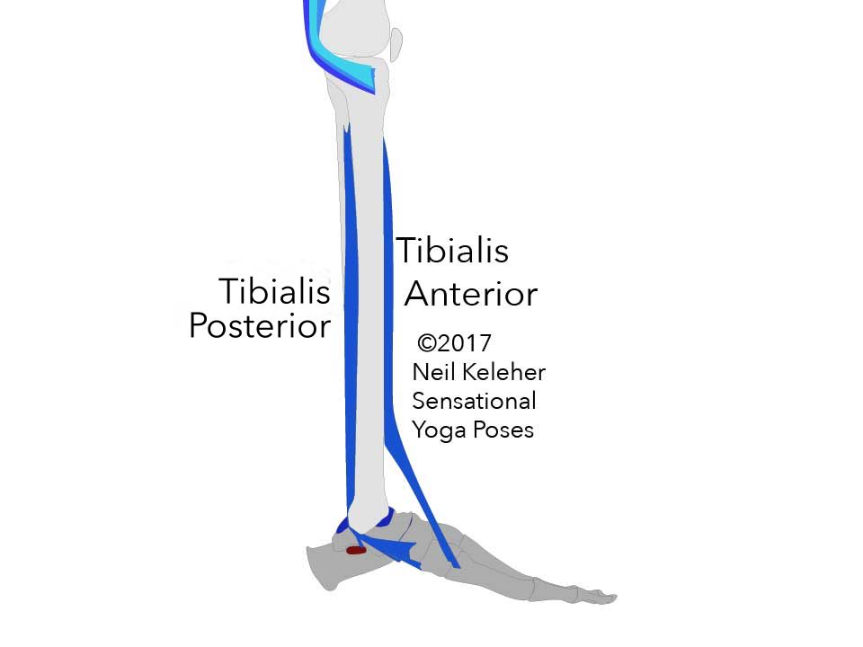
The primary action of rolling the shins out while standing is caused by Tibialis Posterior.
Note that rolling the shins externally can also be caused by the muscles that act on the IT Band.
However, if the focus is on "using the feet" to roll the shins out, then this action can be driven by activation of the Tibialis Posterior.
You may feel tension in the back of your lower leg while rolling your shins out. That could be your tibialis posterior activating.
Tibialis Posterior attaches to the back surfaces of both the fibula and tibia. Its tendon passes down the inside of the ankle to attach to the underside of the inner arch of the foot.
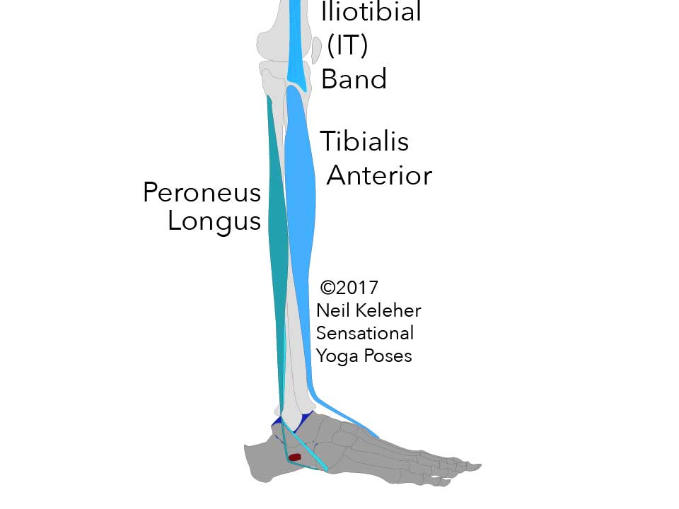
Peroneus longus attaches to the outside of the fibula (the outermost and smallest of the two lower leg bones.)
It passes down the outside of the ankle behind the Lateral Malleolus (the bottom most part of the fibula, that sticks out of the outside of the ankle) and passes under the foot to attach to the inner arch and to the "root" of the big toe.
Because it passes behind and under the trochlear process (peroneal tubercle) on the outside of the calcaneus, Peroneus Longus tends to create a forwards push on the outside edge of the calcaneus which, unresisted, would cause the heel bone to roll inwards. This inward roll of the heel can be countered by an active Tibialis posterior since this muscle has a tendon branch that attaches to the sustentaculum tali on the inside edge of the calcaneus.
Where activation of the tibialias posterior tends to create external rotation of the shin (or a resistance to internal rotation), activation of the peroneus longus (and/or peroneus brevis) can tend to create internal rotation of the shin (or a resistance to external rotation.
Activating together, these two muscles can work against each other to stabilize the shin against rotation while standing.
This can be important for muscles that act on the IT Band as well as the Sartorius, Gracilis and Semitendinosus muscles since these muscles can act from the pelvis to rotate the shin outwards or inwards. With the shin stabilized against rotation by the Tibialis Posterior and Peroneus Longus, these muscles have a stable foundation from which to help control the pelvis.
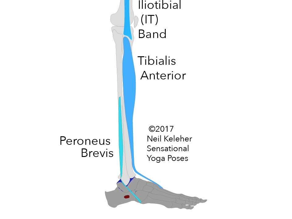
Peroneus Brevis, like Peroneus Longus, attaches outside of the fibula.
It attaches to the lower part of the fibula underneath the peroneus longus.
Activation of the peroneus longus can create a forwards push along the outside of the calcaneus. Meanwhile the peroneus brevis creates a rearwards pull on the outer-most metatarsal.
These opposing actions can work together to help lift the center of the outer arch of the foot.
Tibialis Anterior is situated at the front of the shin.
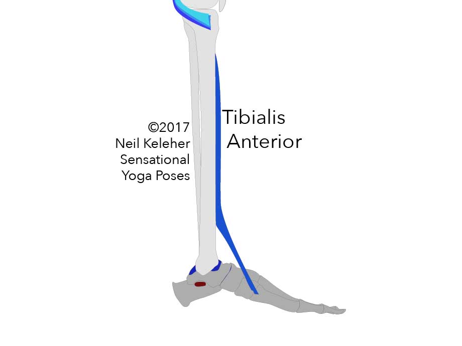
Medial/Inside View
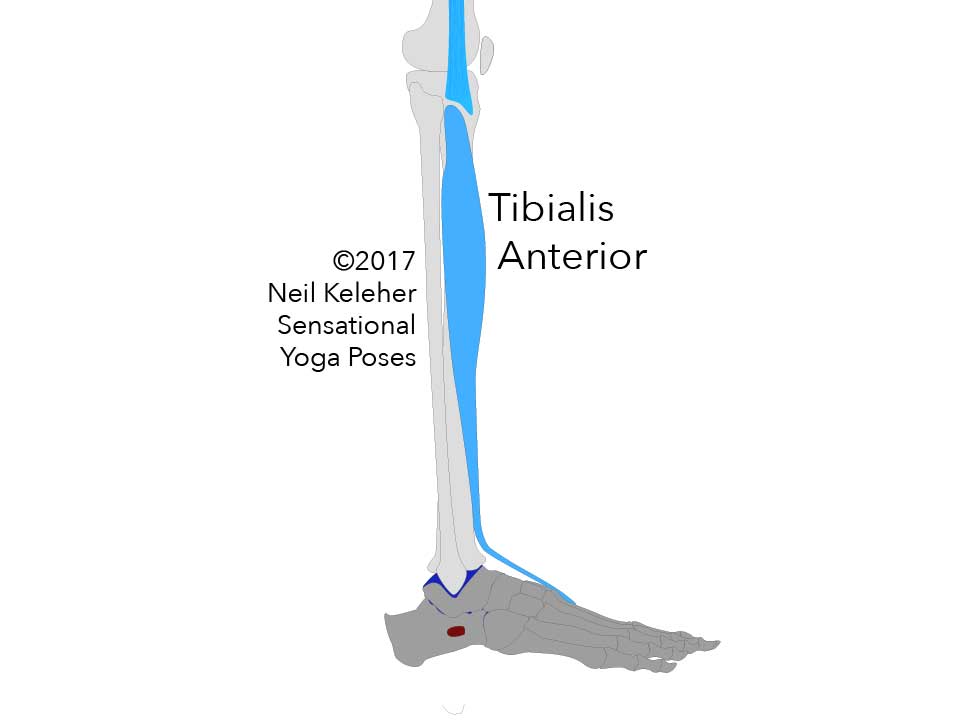
Lateral/Outside View
It attaches to the front of the tibia outside of the tibia, starting high up, where the bone swells to form the bottom of the knee. It passes under the extensor retinaculum, a band of connective tissue at the front of the ankle. From there it runs along the inside edge of the upper surface of the foot to attach to the top surface of the inner most cuneiform and the rearmost part of the top surface of the innermost metatarsal.
From Thomas Meyers Anatomy Trains we know that the Tibialis anterior has a connective tissue connection to the front fibers of the IT band.
(The rear edge of the IT band has connective tissue connections to the Peroneus Longus).
Tension in the Tibialis anterior (and/or the Peroneus Longus) can act as an anchoring mechanism for the IT band so that it it turn can act as an anchor for the Tensor Fascia Latae and/or the Gluteus Maximus.
For myself, on one occasion where I had knee pain pretty much at the spot where the IT band connects to the Tibia, Tibialis Anterior activation helped to alleviate that knee pain.
This sort of muscle activation, activating muscles in the same "anatomy train" is another way of creating stability. Possible names for this mechanism are "Fascial Stability" or "Fascial Anchoring" or to be clearer, "Muscle Activated Connective Tissue Anchoring."
It's basically a means of taking up slack.
It creates stability for a muscle by adding tension to the connective tissue structure that it is a part of.
Published: 2020 08 05
Updated: 2023 03 25
